New Paragraph
Exploring Dental X-Rays: Procedure and Safety Tips
Maintaining good oral health means understanding some important procedures, like dental radiography, often called dental X-rays. These imaging techniques help dentists see the detailed parts of your mouth that you can't see during a regular check-up. X-rays can find early signs of tooth decay. They also help look at the jawbone when planning for dental implants. In this way, X-rays give important information about your oral health.
Understanding Dental X-Rays
Dental X-rays use a small amount of radiation exposure to make images of your teeth, bones, and nearby tissues. This type of medical imaging is significant for finding and treating different oral diseases. X-rays can go through soft tissues, like your gums and cheeks. This allows dentists to see parts of your dental structure that are not visible.
These images help find problems such as cavities between teeth, hidden decay under old fillings, bone loss from gum disease, and how developing teeth are positioned. The information from dental X-rays helps dentists create special treatment plans for each patient.
The Importance of Dental X-Rays
Dental X-rays are very important for finding oral health issues early. They can notice problems before you can see them or feel any signs. This early find helps you avoid bigger and more expensive treatments later.
For example, X-rays can show small cavities forming between teeth. They can also show gum disease with signs of bone loss and infections hiding under the gums. Taking action early can save your natural teeth and help you keep a healthy, bright smile.
Additionally, dental X-rays are needed for treatments like root canals, tooth extractions, and placing dental implants. These images help dentists see clearly, leading to safer and more accurate care.
How Dental X-Rays Work
Dental X-rays work by sending a focused beam of radiation to an area of interest in your mouth. This beam goes through your teeth and jawbone. Different tissues absorb the radiation in varying amounts. Denser tissues, like bones and teeth, absorb more radiation. This makes them show up as white or light gray in the images of your teeth.
On the other hand, softer tissues, such as gums and nerves, absorb less radiation, which results in darker spots on the X-ray. Now, with the latest digital radiography, the amount of radiation exposure has been greatly reduced. These technologies also produce high-quality images.
New tools like cone beam CT scans offer clear three-dimensional images. They are especially helpful for complicated tasks, such as planning for dental implants and performing extractions like wisdom teeth.
Types of Dental X-Rays and Their Uses
Dentists use various kinds of dental X-rays to get a clear picture of your oral health. Each type shows different views and provides special information that helps with diagnosis. When you know what each X-ray is For, you will feel better about your dental care.
Intraoral X-Rays: Detailing the Inside
Intraoral X-rays are exactly what their name implies. They use photographic film or a digital sensor placed inside your mouth. This captures clear images of your teeth and the structures around them. These X-rays are the most common type used during regular dental checkups.
One type of intraoral X-ray is the bitewing X-ray. It focuses on the crowns of your upper and lower teeth while you bite down. This view helps find cavities between teeth and shows changes in bone density that can come with gum disease.
There are other types of intraoral X-rays too. Periapical X-rays show the whole tooth, while occlusal X-rays focus on both the upper and lower jaws. These detailed images are essential for diagnosing many oral health problems.
Extraoral X-Rays: Viewing the Bigger Picture
Extraoral X-rays are different from intraoral X-rays. They use the X-ray film or sensor outside the mouth. These X-rays help get a wider view of the skull, jawbone, and nearby areas.
Panoramic X-rays show a large view of the whole mouth. They include all teeth in both jaws, the jaw joints (TMJ), and the sinuses. This image helps check the overall health of your mouth. It is useful for finding impacted wisdom teeth and looking for problems with the jawbone.
Cephalometric X-rays focus on the side of the head. They give clear images of the jaw compared to the skull. Orthodontists mainly use these X-rays to identify bite issues and create tailored treatment plans for braces and other devices. Advanced imaging methods, like CT scans, are also part of extraoral X-rays. They provide 3D images used in maxillofacial radiology and for planning surgeries.
The Procedure: What to Expect During a Dental X-Ray
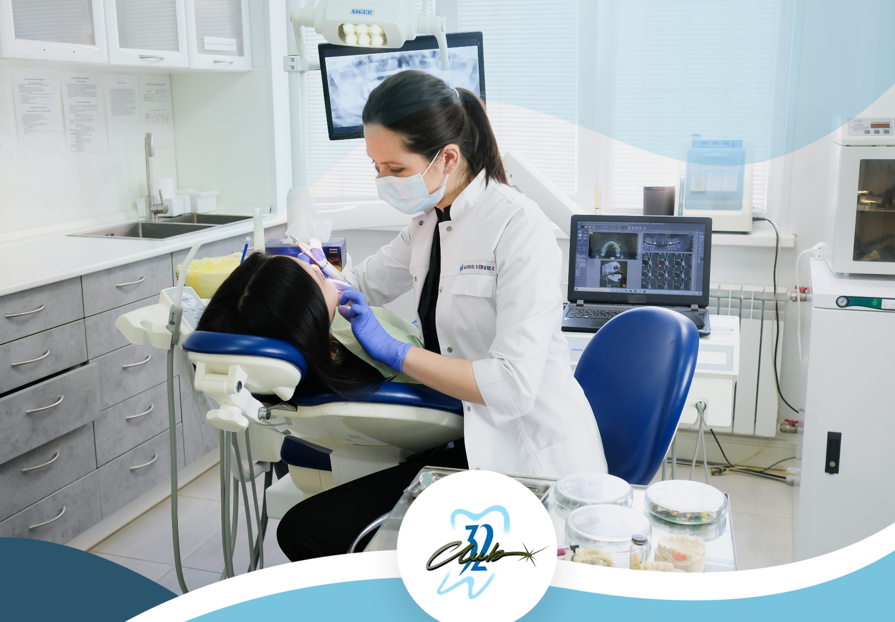
A dental X-ray is a fast and usually painless process. It may feel a little scary, especially if it's your first time, but learning about what happens can ease your worries. Don't worry! Your dentist will help you with every step. They will make sure you are comfortable and feel good during the whole procedure.
Preparing for Your Dental X-Ray
Generally, you do not need special preparation for a dental X-ray. You can eat and take your medication as usual. However, if you are pregnant, it’s important to tell your dentist. They might take extra steps to keep you safe.
When you get to the dentist’s office, especially if you are a new patient, they will ask you about your medical and dental history. This helps the dentist decide what type and how many X-rays you need.
Before the X-ray, you will wear a lead apron over your chest. Sometimes, you'll also wear a thyroid collar. This protects your thyroid gland from radiation exposure. These steps are normal and keep you safe during the process.
Step-by-Step Guide to the X-Ray Process
Once you're prepped and ready, you'll be seated comfortably in the dental chair. Depending on the type of X-ray needed, you'll either bite down on a small plastic piece called a bite blocker or keep your tongue in a specific position to ensure clear images.
| Step | Description |
|---|---|
| 1. Positioning | The dental technician will carefully position the X-ray sensor or film inside your mouth for intraoral X-rays. |
| 2. Adjustment | They will adjust the X-ray machine's arm to align it with the area of interest. |
| 3. Exposure | You'll be asked to remain still while the X-ray is taken, which only lasts a few seconds. |
| 4. Repetition | The process might be repeated a few times to capture different angles or areas. |
Safety Tips and Considerations
Dental X-rays give you a very low dose of radiation. This dose is much less than what you get from natural sources every day. Dentists follow strict safety rules. They use lead aprons, thyroid collars, and smart imaging methods like digital radiography to make sure you get even less radiation.
If you have any concerns about radiation exposure, talk to your dentist. They can answer your questions, explain the safety steps they take, and help you feel safe about the process.
Understanding Radiation Exposure
Dental X-rays use low amounts of radiation. However, over time, this can add up. It is important to keep track of how often you get X-rays and whether they are needed.
The American Dental Association (ADA) has rules for the safe use of radiation in dentistry. Dentists follow these rules closely. They also learn about the latest imaging technology to reduce radiation exposure.
If you choose a new dentist, tell them about your recent X-ray history. This helps them avoid giving you extra X-rays that are not needed. It also helps keep your dental care consistent.
Tips to Minimize Risks
Follow these extra tips to keep yourself safe and reduce risks with dental X-rays:
- Ask about lead apron use: Make sure your dentist gives you a lead apron during X-rays. This apron helps protect you from scattered radiation.
- Inquire about their tools: Choose dental offices that use digital X-rays. They usually emit less radiation than traditional film X-rays.
- Keep a record: Write down your dental X-ray history. Include the dates and types of X-rays you’ve had. This record helps you track your exposure and make smarter choices about future X-rays.
In recent years, dental imaging has become much safer and more effective. By staying informed and talking with your dentist, you can have a safe, comfortable experience. This can help catch oral health issues early and allow for better treatment.
Conclusion
In conclusion, knowing the procedure and safety measures for dental X-rays is very important for your oral health. These imaging methods give helpful details that support accurate diagnosis and treatment planning. By following safety tips and working closely with your dentist, you can have a smooth and safe experience during your dental X-ray. Remember, your dental health is vital. Regular check-ups, including X-rays when needed, help keep your smile healthy. Stay informed, focus on oral hygiene, and trust your dental care team for the best results.
As a leading dental practice in New Jersey,
Club 32 Dentistry is committed to providing exceptional care and state-of-the-art technology.
Our experienced team of dentists and hygienists utilizes advanced X-ray techniques to ensure accurate diagnosis and effective treatment planning. We prioritize patient safety and comfort, and our modern facilities are equipped with the latest safety measures. Trust Club 32 Dentistry for expert dental care and a positive experience.
FAQs
Are dental X-rays safe, and how much radiation is involved?
Yes, dental X-rays are safe and involve a very low dose of radiation—much less than the amount you are exposed to from natural sources daily. Dentists follow strict safety protocols, such as using lead aprons, thyroid collars, and digital radiography, to further minimize radiation exposure. If you have concerns, your dentist can explain the safety measures in place.
How can I reduce my exposure to radiation from dental X-rays?
To reduce your exposure, ensure that your dentist uses a lead apron during X-rays, choose dental offices that use digital X-ray technology (which emits less radiation than traditional film X-rays), and keep a personal record of your X-ray history to avoid unnecessary or duplicate X-rays.
How often should I get dental X-rays?
The frequency of dental X-rays depends on your oral health needs and history. The American Dental Association (ADA) provides guidelines to ensure X-rays are only taken when necessary. It’s important to inform your dentist of any recent X-rays to avoid unnecessary exposure. Your dentist will determine the appropriate schedule for X-rays based on your specific situation.
Need Assistance? We’re Here to Help
Our expert team is ready to support your dental health and well-being.
We are committed to offering personalized dental care solutions that promote a healthy smile.
